-
Li, L., Chen, L. Feldstein, H., Jia, Z., Hu, C., Chen, H., Geng, Y., Peterman, E.M., Slebodnick, C., Speiser, D.I., Baum, D., and Kolle, M., in press at PNAS, A possible biomineralized light guiding structure in the ossicular skeletons of the starfish Protoreaster nodosus
-
Peterman, E.M., Williams, M.L., and Kylander-Clark, A., 2025, Journal of Metamorphic Geology, Vanishing evidence of UHP metamorphism in the Rhodope Metamorphic Complex, Greece. p. 1-19. https://doi.org/10.1111/jmg.70011
-
Johnson, S.A., West, D.P. Jr., and Peterman, E.M., 2023, Geochronology, geochemistry, and tectonic setting of Ordovician metavolcanic rocks in the Liberty-Orrington belt, Maine: Implications for the evolution of peri-Gondwanan arcs in the northern Appalachians, Canadian Journal of Earth Sciences, December, p. 1–18. https://doi.org/10.1139/cjes-2023-0115
-
Hillenbrand, I.W., Williams, M.L., Peterman, E.M., Jercinovic, M.J., and Dietsch, C.W., 2023, Petrochronologic constraints on inverted metamorphism, terrane accretion, thrust stacking, and ductile flow in the Gneiss Dome belt, northern Appalachian orogen, Journal of Metamorphic Geology, p. 1-39. https://doi.org/10.1111/jmg.12741
-
Yang, T., Chen, H., Jia, Z., Deng, Z., Chen, L., Peterman, E.M., Weaver, J.C., and Li, L., 2022, A natural dual-scale single-crystalline architected lattice material. Science, v. 375, issue 6851, p. 647-652. https://doi.org/10.1126/science.abj9472
-
Peterman, E.M., Reddy, S.M., Saxey, D.W., Fougerouse, D., Quadir, Z., and Jercinovic, M.J., 2021, Trace element segregation to dislocation loops in experimentally heated zircon. American Mineralogist, v. 106, no. 12, 1971-1979. 10.2138/am-2021-7654
-
West, D.P. Jr., Peterman, E.M., and *Chen, J., 2021 Silurian-Devonian Tectonic Evolution of Mid-Coastal Maine, U.S.A.: Details of Polyphase Orogenic Processes. American Journal of Science, v. 321, p. 458–489. https://doi.org/10.2475/04.2021.03
-
Peterman, E.M., Jercinovic, M.J., Beane, R.J., and *de Wet, C.B., 2021, Kyanite preserves prograde and retrograde metamorphic events as revealed by cathodoluminescence, geochemistry and crystallographic orientation. Journal of Metamorphic Geology, p. 1-24. https://doi.org/10.1111/jmg.12593
-
Peterman, E.M., Reddy, S.M., Saxey, D.W., Snoeyenbos, D.R., Rickard, W.D.A., and Fougerouse, D., 2019, Nanoscale processes of trace element mobility in metamorphosed zircon, Contributions to Mineralogy and Petrology, v. 174, no. 92, 29 pp. https://doi.org/10.1007/s00410-019-1631-1
-
Sullivan, W.A., and Peterman, E.M., 2017, Pulverized granite at the brittle-ductile transition: An example from the Kellyland fault zone, eastern Maine, U.S.A. Journal of Structural Geology, v. 101, p. 109–123. http://dx.doi.org/10.1016/j.jsg.2017.07.002
-
Peterman, E.M., Reddy, S.M, Saxey, D.W., Snoeyenbos, D.R., Rickard, W.D.A., Fougerouse, D., and Kylander-Clark, A.R.C., 2016, Nanogeochronology of discordant zircon measured by atom probe microscopy of Pb-enriched dislocation loops. Science Advances, v. 2, e:1601218, 9 pages. doi: 10.1126/sciadv.1601318
-
Peterman, E.M., Snoeyenbos, D.R., Jercinovic, M.J., and Kylander-Clark, A., 2016, Dissolution-reprecipitation metasomatism and growth of zircon within phosphatic garnet in metapelites from western Massachusetts. American Mineralogist, v. 101, p. 1792-1806. https://doi.org/10.2138/am-2016-5524
Scanning Electron Microscope (SEM)
Uses
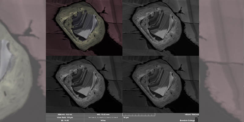
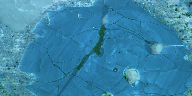
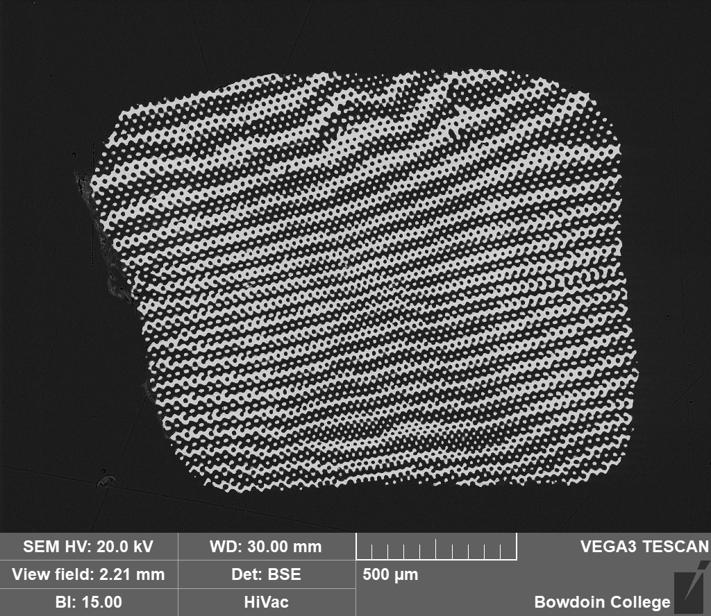
A scanning electron microscope (SEM) allows one to see well beyond the powers of the naked eye. It can, for instance, provide details or reveal history about the composition and formation of rocks, minerals, clamshells, corals, and ancient coins.
Features
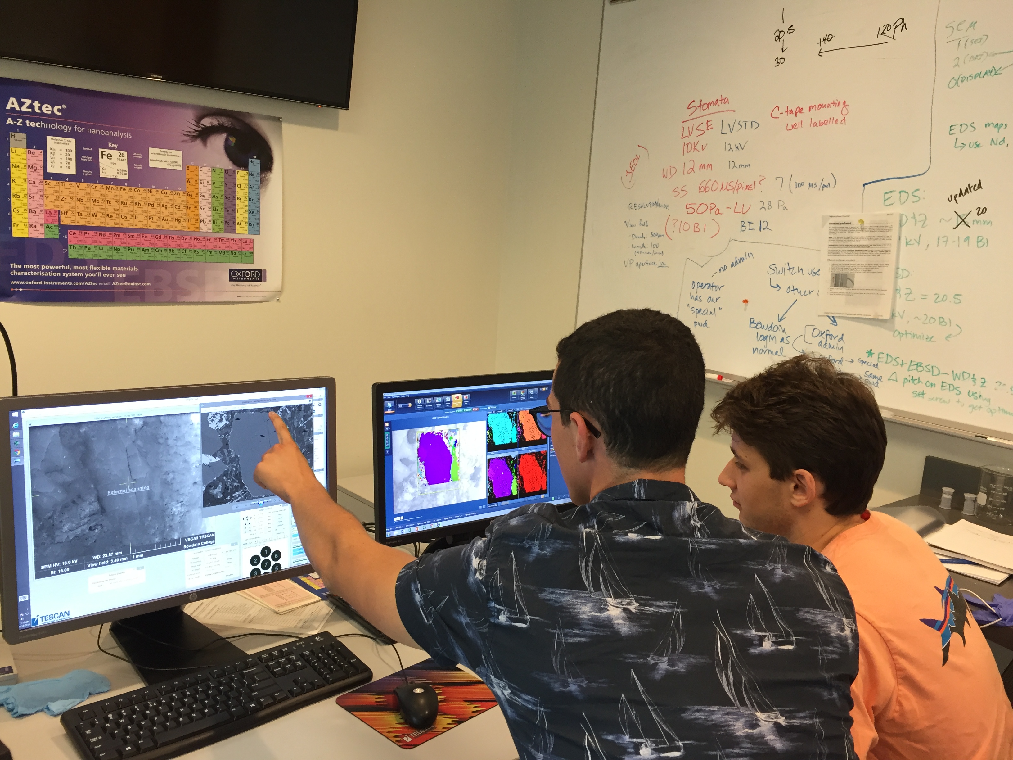
The Tescan Vega3 LMU Scanning Electron Microscope (SEM) is equipped with an array of detectors. The backscattered electron (BSE) and cathodoluminescence (CL) detectors image samples, and the energy dispersive spectrometer (EDS) and electron backscatter diffraction (EBSD) systems collect quantitative data on chemical compositions and crystal orientations. The SEM is located in the Roux Center for the Environment.
The personal-computer controlled instrument allows:
- Digital imaging (up to 300,000x magnification at c. 1 nm resolution)
- Chemical analysis
- Crystal orientation mapping
Using the SEM
The SEM is used by students and faculty throughout the College. A range of specimens may be imaged and analyzed, including minerals, sea urchin spines, microfossils, nematodes, plants, bone tools, coins, and ceramics.
The instrumentation facilitates innovative research that involves professors, collaborators, students, and community partners and expands the breadth of scientific inquiry possible at the College.
The new instrumentation also provides excellent opportunities for undergraduate research training. Undergraduates use the SEM in multiple courses, as well as during independent study and summer research projects. These research projects train students to apply modern analytical techniques to original research questions and enhance the infrastructure for research across disciplines at the College.