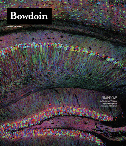
The Brainbow Connection
By Michael Blanding for Bowdoin MagazineJeff Lichtman ’73 was a kid who wanted to look at everything, to uncover secrets he knew even the most ordinary of objects around him held.
As a Harvard neuroscientist and researcher, Lichtman continues this dedication to close looking through his quest to map the brain in its infinite complexity.
As dean, he wants every science student to feel like an explorer on a new continent, to wonder what it is that they might see.

Lichtman in his lab. To his right and behind him are multibeam electron microscopes that use ninety-one parallel beams to create the imaging speed necessary to process the enormous datasets they use. To his left is a new gas cluster milling system that allows them to generate image stacks with slices as thin as what you would get if you cut a single sheet of paper into 5,000 ultra-thin sheets.
“Think of an American flag,” says neuroscientist and Harvard professor of molecular and cellular biology Jeff Lichtman ’73, sitting in the office at his lab. In response, he says, you might envision a field of blue, speckled with fifty bright stars and laced with thirteen red and white stripes. “You’d have no trouble rendering that, and it doesn’t take a week—you render it instantaneously,” says Lichtman. “I could have asked you to render your mother, or your pet dog, or your child—it doesn’t matter, it’s there immediately.”
Easy—but here’s the hard part: Where in the brain is that memory sitting? “Is it a physical entity? Does it weigh something?” Lichtman asks. “It has to be something so stable that even if you’re not using that memory—sometimes for decades—it’s still there, just like it was yesterday.” We might imagine a vast file cabinet in the brain, with drawers labeled “American flag” and “mother” and files made up of proteins or other material waiting to be retrieved.
Lichtman has a different theory, on the forefront of brain science: that everything we know and learn is embedded not into any particular molecule, but rather in the connections between nerve cells. “Each memory is not a chemical, it’s a pathway through this vast morass of wires,” he says. “What’s being triggered is a movement of information through that wiring system in the brain.” While the Human Genome Project has mapped the genes in the body and examined their functions, Lichtman believes that when it comes to the brain, genes are not as important as cells and how they are connected.
He’s spearheaded an ambitious project to map the brain that he’s dubbed the “connectome,” tracing the millions of miles of neurons packed into that relatively small space to understand how people learn and remember, as well as why sometimes the wiring goes wrong. Last year, Lichtman was appointed dean of science for Harvard’s Faculty of Arts and Sciences. A week later, he and his lab colleagues published a landmark paper in Science announcing the largest image of the brain to date: a digital three-dimensional rendering of a cubic millimeter of human brain tissue that contains 57,000 cells with 150 million synapses between them—1,400 terabytes of data in all.
The scale of the image is staggering. “Words fail us, not because we’re so emotionally overcome, but because the complexity of the wiring diagram cannot be put into sentences,” he says. “It’s like trying to describe all of New York City—even if you had a complete map of every single thing going on, words would not be able to capture it.” To overcome those limitations, Lichtman has developed innovative techniques to depict the brain visually, including a collaboration with Google using artificial intelligence to color tiny cells and parts of cells within. Already, the techniques have revealed never-before-seen structures with the potential to revolutionize brain science. There’s no telling what he might find on the way to his ultimate goal of mapping the complexity of the brain in its entirety.

A Brainbow image of the axon tracts in the brainstem of a mouse.
Sculpting the Mind
When Lichtman was a kid growing up in Westchester, New York, his father did something early on that transformed his son’s life. A hematologist at a nearby hospital, he placed his old Leica medical-school microscope in the bedroom Lichtman shared with his brother. “I don’t remember him giving us any instructions, but I used it to look at everything—throughout my childhood, there was no aspect that wasn’t scrutinized.” He remembers keeping a Tupperware container of smelly pond water in the bedroom, spending hours watching paramecia and other protozoa swimming around “a whole gigantic world.”
Even now, a microscope sits on the desk in his sunny office, and Lichtman uses it regularly to illustrate points to students. “I actually can’t imagine an office without a microscope.” At Bowdoin, he aspired at first to be a writer or musician, but science was effortless when those subjects were not. “Biology was just a natural for me,” he says. “The advice I give to young people now is to choose what you find easiest—because if you keep pushing on what you find easy, you are going to get to a point where you find it hard, and by then, not many people will be at your level.”
Lichtman took his own advice, going into an MD/PhD program at Washington University in Saint Louis. Still, the liberal arts environment in his undergraduate years not only shaped his career but also influenced his thinking. “I give Bowdoin a lot of credit for encouraging that liberal arts worldview, where you are really not a complete human being if all you do is advanced math or electronics,” he says, recalling evenings at professors’ houses and long conversations over dinner that blurred the line between classroom and life.

“He is a true philosopher of science,” says Bobby Kasthuri, a former student at Washington University and longtime collaborator.
“Jeff is a natural contrarian, but there’s a kind of gentleness to his contrarian nature. He’ll talk about radical ideas but in a laid-back fashion.”
With the rise of molecular biology, neuroscientists increasingly looked to explain cellular behavior through genes and the molecules they produce, developing a theory that pathways were strengthened in the brain through the transmission of chemicals between synapses. Lichtman, by contrast, developed a belief that it wasn’t chemicals as much as the physical structure of the brain that determines its function. “Jeff has long argued that it’s very hard to go from molecules to behavior,” says Kasthuri. “His argument is that cells are the fundamental units. The way they connect is what really matters.” That view reshapes how we think about the brain and how it develops. Most people assume that our brains get more complex as we age, accumulating connections, but Lichtman sees it differently. “You are not building a wiring system as you get older, you are sculpting one,” he says. “You start out with wires for everything, and you ultimately keep the things you learn and get rid of the rest.”
As we prune connections, he says, older people literally get set in their ways. “They would call it wisdom,” he says. “You might call it being stuck in the past.” The process carries broad implications—psychologically, sociologically, even politically. “Different adults can see the same thing in radically different ways,” Lichtman says. “That is because they’ve developed different pathways based on what they’ve read, heard, and thought about. It becomes very hard to change that.” On the other hand, he says, humans’ ability to change their brain wiring has made them uniquely resilient and adaptable as a species. “There was a time when humans didn’t live indoors or wear clothes,” Lichtman says. “We certainly didn’t read or write. Now we are not only able to encode information in our brains but also pass it along to others. That means humans keep changing because of this profound ability to adapt our wiring diagrams to the world we live in.”

Neurons colored to differentiate size and to show layers. Large neurons are colored red and small ones blue.
The same adaptability can also have a darker side, helping explain some forms of mental illness. Children raised in a dysfunctional or abusive home might adapt in ways that lead to emotional and behavioral problems or addiction. Other diseases, such as Alzheimer’s and epilepsy, seem to have a genetic component as well. Decades of attempts to pin those conditions on a single gene, or even a combination of genes, have failed. Lichtman speculates that in reality the causes are more complicated, stemming from problems that grow out of a miswiring in the diagram of the brain. “And until now, seeing that diagram has been impossible.”
That conviction leads him back to a principle that has influenced him since his childhood pond-water watching: the power of observation. Science is always performed through hypothesis and experimentation, he says. Whether it’s astronomers charting galaxies, particle physicists smashing atoms, or Darwin deriving evolution from finches’ beaks, sometimes the most fundamental discoveries arise from simply seeing what’s out there.
If we want to cure diseases of the brain, first we must understand what the brain looks like—just like someone would need to know the way a car works to fix an oil leak or a wiring diagram to fix an electrical system. “We’re just starting to look at these wiring diagrams, and already we’re seeing things that aren’t in any textbook, just because no one has had a way to look for them before.”

A section of mouse brain cortex showing a large dendrite (red) with axons and synapses—the points where signals pass from one neuron to another.
A Rainbow of Neurons
Lichtman first started tracing these neuronal connections at Washington University, looking at clusters of nerves in the peripheral nervous system called the autonomic ganglia, which control subconscious functions such as sweating, goosebumps, or salivation. He laughs while recalling how he explained his dissertation work to his mother. “I told her I found this huge reorganization of the writing diagram of the salivary gland. She said, ‘What the hell—you went to school so you can study saliva in rats?’”
Beyond those specific structures, however, Lichtman was making groundbreaking progress in mapping how nerves connect, laying the groundwork for the new field of connectomics. After earning his MD/PhD in 1980, he became a professor at Washington for two and a half decades before moving to Harvard in 2004. At first, he used physiological techniques to stimulate nerve cells to determine which were connected. Nerve cells, or neurons, consist of a long spine called a dendrite, ending in a spiky structure called an axon, which connects to other nerve cells across small gaps called synapses.
Lichtman realized he could more elegantly trace those connections using color. Traditionally, scientists stained a few cells at random, hoping to glimpse a connection. Stain too many, and the result was a blurry mess. Lichtman had a different idea. Using a recently discovered gene from jellyfish that produced fluorescent proteins, he realized he could manipulate it to create red, green, and blue proteins in mouse cells. By inserting those genes into cells and randomly exciting or inhibiting them, he could produce a spectrum of colors from proportions of those three colors, similar to the way a color television creates a rainbow of color through three colors of backlit pixels. He called it the “brainbow.”
Under black light, the neurons glowed in a riot of colors, each distinguishable from their neighbors under the microscope. Publishing the results in an article in Nature in 2007 that coined the word “connectomics” for the first time, Lichtman was able to showcase images of the brain that were not only informative, but also stunningly beautiful, resembling a lit-up fiber-optic Christmas tree.
While the technique worked well for the peripheral nervous system, the random colors failed to provide the resolution necessary in the densely packed central nervous system. To make sense of that tangle, Litchman and his colleagues would have to use a different approach. From his office, he leads the way down an elevator to the basement, where he shows off a machine that looks like a film projector called the automatic tape-collecting lathe ultramicrotome (ATLUM). The device, Lichtman explains, uses a diamond knife to cut sections of brain thirty nanometers thick—a thousand times thinner than a human hair.

A pyramidal neuron from a human brain sample, with axons showing the rare whorl pattern. Of the millions of axons in the data set processed by Lichtman’s lab, only a few had these whorls.
Those sections are collected on a silicon wafer, which can be imaged separately by an electron microscope and then recollected digitally to create a three-dimensional section of tissue. After digitizing the brain slices, the scientists collaborated with Google, which worked to stitch those black-and-white images into a seamless three-dimensional representation of the brain. Using artificial intelligence, technicians have been able to trace individual neurons and color them with enough differentiation to distinguish not only individual cells, but also parts within cells.
At the pace of traditional instruments, Lichtman calculated that the tens of thousands of images for a cubic millimeter of brain would take seventeen years to complete. So, he and his colleagues built faster systems, including an electron microscope the size of a refrigerator that can beam a shower of sixty-one streams of electrons at a time, reducing the time necessary to six months. They are now working on a new machine that will image ninety-one images at a time, connected to a device with a tangle of parts and wires that will automatically shave pieces of brain and feed them into the machine without need for human interaction.
The amount of computing power to accomplish the task has been so vast that Google has had to develop entirely new software to allow multiple computers to work on the task at once. Google research scientist Viren Jain compares it to technological innovations produced by NASA as it has worked to solve problems of spaceflight. “There are these kinds of grand problems, where if you choose something hard enough, you end up inventing something that is more broadly useful.”
Back in his office, Lichtman shows off the results as he zooms on his computer monitor through a vast virtual landscape of colored wires that represents a tiny section of the cerebral cortex of a forty-five-year-old woman who was undergoing neurosurgery for epilepsy. Within that, he and his fellow researchers have discovered previously unknown brain structures. In one, which he has dubbed “whorls,” axons look like a mass of spaghetti all twisted up. It’s the kind of thing that could only be discovered in a dataset this enormous. “Very few axons in the dataset are doing this—maybe twenty out of 200 million—so it’s very rare,” says Lichtman, who speculates it might be related in some way to epilepsy, potentially serving as a marker for the disease.
In another structure he calls a “superconnection,” a nerve cell makes dozens of connections to the same cell. “For most connections, all it takes it one dendritic spine passing by a cell to create a synapse that can carry information,” Lichtman explains, zooming in to show a colored wire that connects to a cell in dozens of places, going up one side and down the other. “Instead of making one synapse, it’s making fifty-two.” He hypothesizes that these strong connections might represent firmly rooted learning, where a concept becomes automatic— the way we might instantly imagine a man with a stovepipe hat and beard from the single word “Lincoln,” or step on the brake as soon as we see a red light while driving. “You don’t have to think, ‘I have to take my foot off the gas and put it on a brake pedal,’” he says. “It’s an automatic fast path through the brain.” For Lichtman, finding new structures like this is a thrill akin to discovering new species of animals in an unknown land. “Suddenly there are all these birds and reptiles you’ve never seen before,” he says. “That’s how it feels, like an explorer arriving on a new continent.”
Tackling GHPS
As Harvard’s new dean of science, Lichtman has been working to re-create that same spirit of exploration for students, rethinking how to approach scientific inquiry in an age of the internet and artificial intelligence. “One of the crises of science teaching is that all the information known in the world is available at the fingertips of every student with a cell phone,” he says. “I wanted to come up with a program where ChatGPT would be useless.”
The solution he came up with is a new program based on what he calls GHPs—genuinely hard problems—including the physical roots of mental illness, the nature of dark matter, and the solution to climate change. “These are problems where, despite the best efforts of modern science, we’ve made no progress,” he says. “So, students are not at as much of a disadvantage as you might think, because all the people who know everything still haven’t gotten it right.”
Students would take a class their first semester on campus in which faculty present a number of these problems, he says, and then they’d pick a faculty member who would suggest courses they could take and books and papers they could read to try to solve it over the next four years. “If they are successful, they get a Nobel Prize,” Lichtman quips. More to the point, however, they’ll get something even more essential for a budding scientist: training in a way of thinking to confront the unknown with excitement over the possibility of discovering something new.
“Most education in the sciences consists of courses in which someone tells you, ‘Trust me, you are going to need this later in life,’” he says. “But it doesn’t serve them well when we give them a bunch of things to memorize that they could get off the internet. Most of our students have gotten into Harvard by focusing on accomplishment, but that’s different than battling a very hard problem, where you might fail many times before succeeding.”
The prime attribute he’s hoping the program engenders in students is curiosity. “Good scientists are confronting problems every day that they don’t have the answers to,” he says. “Every night they are thinking about them, and every morning, they wake up still thinking about them—they just can’t help it.”
He might just as well be talking about himself, as he works to expand on the small piece of brain he’s already helped image. With the new microscope system at Harvard, along with a similar one at Princeton, he thinks he and his collaborators can image a piece of brain ten times larger than the one in their Science paper in just two years. “If we had twenty-five of these instruments, we could do a whole mouse brain in two years,” he says. Of course, that would require massive amounts of data analysis and storage, on the order of exabytes—that is, millions of terabytes—but the payoff could be incredible. “No mammal’s wiring system is known at all,” he says. “Once we had a normal mouse, we could then do a mutant animal.”
By comparing the two, neuroscientists could potentially understand brain disease in an entirely new way, developing therapies to target the connections in the brain the same way that the Human Genome Project has led to innovative new molecular therapies. The truth, Lichtman says, is that no one knows exactly what they’ll find, what new structures await discovery in the microscopic depths of the human brain, and how they might transform our understanding of learning, memory, and illness.
“People who do this work consider themselves explorers,” Lichtman says again. “We just want to see what’s out there.” On his desk, the microscope awaits—as it has for all his life—a reminder that discovery begins not with answers, but with a willingness to look closely at the world.
Michael Blanding is a Boston-based investigative journalist whose work has appeared in The New York Times, WIRED, Smithsonian, Slate, The Nation, The Boston Globe Magazine, and Boston.
Jared Leeds is a lifestyle, portrait, and documentary photographer based in Boston.
This story first appeared in the Fall 2025 issue of Bowdoin Magazine. Manage your subscription and see other stories from the magazine on the Bowdoin Magazine website.