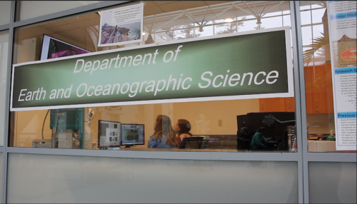
Over January break, geologists Rachel Beane and Emily Peterman oversaw the careful installation of Bowdoin’s new $437,000 scanning electron microscope in Druckenmiller Hall.
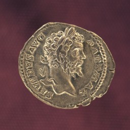
The rather modest size of the microscope, which looks a bit like a desktop computer tower, belies its incredible power to see well beyond the powers of the naked eye. It can, for instance, provide details about the composition and chemistry of samples from rocks to Roman coins, offering a peek into the history behind the objects and how they were formed.
The microscope, which was purchased with a grant from the National Science Foundation, replaces an older one bought in 1999 that was growing less reliable as the years wore on. The new microscope can do more quickly what the old one did. It also has additional capabilities.
Beane and Peterman, who both teach in Bowdoin’s Earth and Oceanographic Science department, will use the new microscope to analyze minerals to learn more about geologic and tectonic processes. Associate Professor of Classics Jim Higginbotham uses the microscope to analyze some of the Bowdoin College Museum of Art’s 2,000 ancient coins. And archaeologist Genevieve LeMoine, curator of the Arctic Museum, employs the microscope to glean information about the provenance of iron tools crafted by the prehistoric Dorset people of the high Arctic.
Electron microscopes work by sending a beam of electrons down a long vacuum. When the beam makes contact with the sample, the electrons interact with the atoms on the surface of the sample. This interplay of atoms can convey much useful data. With six detectors that perform different functions, the electron microscope offers a wide range of information about any number of objects — a shell, a piece of hair, a leaf, a fly’s egg, etc. Samples do not get destroyed in the process.
Some of the detectors, for example, produce images that geologists can decipher to learn the conditions (e.g., temperature, pressure and depth) under which minerals formed. Beane specifically uses electron microscopes to study supervolcanoes. Peterman analyzes rocks that have traveled into the earth’s crust and mantle and back to the surface again to figure out how fast the rocks traveled, what happened to them below the earth’s surface and why they returned. “For the rocks I am studying, we know a lot about how they came back up to the surface, but we don’t know as much about their earlier history because mineral reactions overprinted that history on the way back,” she said.
LeMoine uses the electron microscope to determine the nickel content in prehistoric iron tools she has excavated from Arctic sites. The amount of nickel in a knife or harpoon head made by the Dorset people can tell her whether the iron came from the remnants of an enormous meteorite that crashed into Greenland between 5,000 and 10,000 years ago, or from the Vikings. In this way, she can track the contact between the two groups of early peoples.
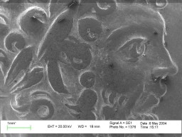
Higginbotham can use the detectors to study the metallurgy of coins. For example, he can look at silver coins minted by Mark Antony (83 BCE – 30 BCE) in an electron microscope to determine the amount of silver in each piece compared to say, lead. This ratio offers a sense of the economic stress Antony was under during his battle with Octavian. As their fight waged on, Antony kept producing more coins to pay his military, and so had to be sparing with his silver. The microscope also produces images that allow Higginbotham to see the artistic details of coins and how much the money was circulated.